File:Hepatitis B virus v2 (3).svg

File originale (file in formato SVG, dimensioni nominali 512 × 363 pixel, dimensione del file: 142 KB)
| Questo file proviene da Wikimedia Commons. La pagina di descrizione associata è riportata qui sotto. |
Indice
Dettagli
| DescrizioneHepatitis B virus v2 (3).svg |
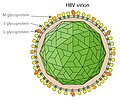
English: Simplified drawing of the Hepatitis B virus particle and surface (surplus) antigen. Created by en:User:GrahamColm |
|||
| Data | 14 novembre 2007 (data di caricamento originaria) | |||
| Fonte | Transferred from en.wikipedia | |||
| Autore | Original uploader was TimVickers at en.wikipedia | |||
| Licenza (Riusare questo file) |
Released into the public domain (by the author). | |||
| Altre versioni |
[modifica] |
|||
| SVG sviluppo InfoField | Questa grafica vettoriale è stata creata con Inkscape.
|
Extended description
The structure of the Hepatitis B virus as first described by Dane & al.[1] and Jokelainen, Krohn & al.[2] in 1970:
Virion
The hepatitis B virion, is a complex, spherical, double shelled particle with a diameter of 42 nm.[1][2][3]
- The 6 nm[2] thick outer viral envelope or membrane contains host-derived lipids and surface proteins,[2] known collectively as HBsAg.[3] The membrane contains globular subunits each measuring ca. 3 to 4 nm in diameter and 3 to 4 nm apart.[2]
- Within the membrane sphere is a 2 nm thick icosahedral nucleocapsid inner core composed of protein (HBcAg) with a diameter of 27 nm.[2] When viewed through an electron microscope the inner core may appear pentagonal or hexagonal,[2] depending on the relative position of the sample.
- The nucleocapsid contains a viral genome[2] of circular, partially double stranded DNA[3] and endogenous DNA polymerase[4][3] within a diameter of ca. 18 nm.[2]
The virion was initially referred to as the Dane particle.[4] Only after Baruch Blumberg received the Nobel Prize in Medicine in 1976 was it universally accepted that the particle is a virus and the infectious agent of Hepatitis B.
Australia antigen (HBsAg)
The serum of infected patients also contain small spherical and rod-shaped particles with a diameter of ca. 20 nm,[5] consisting of surplus virus-coat material containing the HBsAg antigen.[1][2] This antigen was first discovered by Baruch Blumberg in 1965 in the blood of Australian aboriginal people and initially known as "Australia antigen".[6] It was shown to be associated with "serum hepatitis" by A. M. Prince in 1968.[7]
The outer membrane of the virion is sometimes extended as a tubular tail on one side of the virus particle (not shown);[2][3] these virion "tails" are identical to the small particles.[2][3]
The hepatitis B e antigens (shown) are considered not part not part of the viral particle.
References
- ↑ a b c D.S. Dane , C.H. Cameron , Moya Briggs (1970). "Virus-Like Particles in Serum of Patients with Australia-Antigen-Associated Hepatitis". The Lancet 295: 695–698. DOI:10.1016/S0140-6736(70)90926-8.
- ↑ a b c d e f g h i j k l P. T. Jokelainen, Kai Krohn, A. M. Prince and N. D. C. Finlayson (1970). "Electron Microscopic Observations on Virus-Like Particles Associated with SH Antigen". J Virol. 6 (5): 685-689. ISSN 1098-5514.
- ↑ a b c d e f The hepatitis B virus. WHO.
- ↑ a b Almeida J D, Rubenstein D & Scott E J. (1971). "New antigen-antibody system in Australia-antigen-positive hepatitis". The Lancet 298 (7736): 1225–7. DOI:10.1016/S0140-6736(71)90543-5.
- ↑ Bayer, M. E., B. S. Blumberg, and B. Werner (1968). "Particles associated with Australia antigen in the sera of patients with leukemia, Down's syndrome and hepatitis.". Nature (London) 218: 1057-1059.
- ↑ Baruch S. Blumberg, Harvey J. Alter, and Sam Visnich (Jul 1984). "Landmark article Feb 15, 1965: A 'new' antigen in leukemia sera. By Baruch S. Blumberg, Harvey J. Alter, and Sam Visnich". JAMA 252 (2): 252–7. DOI:10.1001/jama.252.2.252. PMID 6374187. ISSN 0098-7484.
- ↑ Prince, A. M. (1968). "An antigen detected in the blood during the incubation period of serum hepatitis". Proc. Nat. Acad. Sci. U.S.A. 60: 814-821.
Licenza
| Public domainPublic domainfalsefalse |
| |
Questa immagine è stata rilasciata nel pubblico dominio dal suo autore, TimVickers nel progetto inglese Wikimedia Commons. Questa norma si applica in tutto il mondo. Nel caso in cui questo non sia legalmente possibile: |
Registro originale del caricamento
- 2007-11-14 18:14 TimVickers 843×577× (81917 bytes) Simplified drawing of the Hepatitis B virus particle and surface (surplus) antigen
Didascalie
Elementi ritratti in questo file
raffigura
14 nov 2007
image/svg+xml
Cronologia del file
Fare clic su un gruppo data/ora per vedere il file come si presentava nel momento indicato.
| Data/Ora | Miniatura | Dimensioni | Utente | Commento | |
|---|---|---|---|---|---|
| attuale | 16:35, 2 ago 2024 |  | 512 × 363 (142 KB) | Glrx | crop with viewBox |
| 20:35, 23 gen 2013 |  | 744 × 1 052 (142 KB) | Graham Beards | More accurate location of core |
Utilizzo del file
La seguente pagina usa questo file:
Metadati
Questo file contiene informazioni aggiuntive, probabilmente aggiunte dalla fotocamera o dallo scanner usati per crearlo o digitalizzarlo. Se il file è stato modificato, alcuni dettagli potrebbero non corrispondere alla realtà.
| Titolo breve | HBV |
|---|---|
| Larghezza | 100% |
| Altezza | 100% |


